Lumbar Facet Joint Disorders: Symptoms, Diagnosis, and When to Seek Care
Lumbar facet joint disorders are a common cause of low back pain, especially when pain feels worse with standing upright, leaning backward, or twisting. The discomfort is often centered in the lower back, but it can also spread into the buttocks or upper thigh. Because many back conditions can feel similar, diagnosis usually combines symptoms, a physical exam, and sometimes imaging or diagnostic injections.
This article explains what lumbar facet joint pain commonly feels like, how it is evaluated, and what to watch for if your symptoms may be coming from something more serious than the facet joints alone.
What are the lumbar facet joints?
The facet joints are small joints at the back of the spine that help guide movement and provide stability. In the lower back, they help control bending, extension, and rotation. When these joints become irritated by arthritis, inflammation, injury, joint degeneration, or excess loading, they can become a source of ongoing pain and stiffness.
Common symptoms of lumbar facet joint disorders
Facet-related pain is usually described as a dull, aching low back pain with stiffness. It may come on gradually or flare after repetitive strain, prolonged standing, awkward posture, or spinal extension.
Common symptoms include:
- Localized low back pain near one or both sides of the spine
- Morning stiffness or pain after long periods of inactivity
- Pain with extension or rotation, such as leaning back, twisting, or standing for long periods
- Relief with changing position, gentle movement, or sometimes bending slightly forward
- Tenderness over the affected area
- Referred pain into the buttocks, hip area, or upper thigh
Some people also notice a grinding or catching sensation during movement, especially when there is more advanced joint wear.
Can lumbar facet pain cause leg pain?
It can, but the pattern matters. Facet joint pain may refer discomfort into the buttocks or thighs, yet it usually does not travel far below the knee in a classic nerve-root pattern. If pain shoots sharply down the leg, causes numbness, tingling, weakness, or is accompanied by reflex changes, a clinician may look more closely for nerve compression, disc problems, or another cause of sciatica-like symptoms.
How facet joint pain is different from other back pain
One reason lumbar facet disorders are hard to identify is that their symptoms overlap with other low back conditions.
- Facet joint pain often worsens with spinal extension, twisting, and prolonged standing.
- Disc-related pain may be more aggravated by forward bending, coughing, or sitting for long periods.
- Sacroiliac joint pain may feel lower and more off to one side near the pelvis.
- Nerve root irritation is more likely when pain is electric, burning, or associated with numbness or weakness.
Because symptom patterns can overlap, a diagnosis should not rely on location alone.
How lumbar facet joint disorders are diagnosed
Diagnosis usually happens in stages, starting with the simplest and least invasive steps.
1. Medical history
A clinician will usually ask about:
- When the pain started
- Whether it came on after injury, overuse, or gradually over time
- What movements make it worse
- What gives relief
- Whether symptoms spread into the leg
- Past back issues, arthritis, or prior treatments
2. Physical examination
The exam may include posture, gait, spinal motion, and palpation of the lower back. Pain reproduced by extension and rotation can raise suspicion for facet involvement, but it is not enough on its own to confirm the diagnosis. A neurological exam is also important to check strength, sensation, and reflexes.
3. Imaging when needed
Imaging may help rule out other causes or show signs that support the diagnosis:
- X-rays may show joint space narrowing, bone spurs, or degenerative changes.
- CT scans provide clearer detail of bone structures and arthritic changes.
- MRI is useful when soft tissue, nerve involvement, swelling, or another source of pain is suspected.
- SPECT/CT may sometimes be used in more complex cases to look for metabolically active inflammation.
It is important to know that imaging findings do not always match symptoms. Some people have degenerative changes on scans without significant pain, while others have pain that is more obvious clinically than radiologically.
4. Diagnostic medial branch or facet joint blocks
When the diagnosis remains uncertain, a specialist may use a fluoroscopy-guided local anesthetic injection to test whether the facet joint or its nerve supply is the likely pain source. If pain drops significantly during the anesthetic window, it can support the diagnosis. Because false positives can happen, some clinicians use controlled or repeat blocks before moving toward more targeted procedures.
When conservative care is usually tried first
Many people with suspected lumbar facet pain start with conservative treatment before advanced procedures are considered. Depending on the individual case, this may include:
- Activity modification during flares
- Physical therapy focused on mobility, trunk control, and core support
- Anti-inflammatory medication if appropriate and approved by a clinician
- Heat or ice for short-term comfort
- Posture and lifting adjustments during daily activities
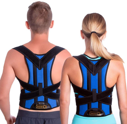
For some people, external support can also be helpful during short periods of aggravation or when daily tasks repeatedly stress the lower back. If you are looking for added support during conservative management, you can explore this adjustable lumbar support brace as a practical option for compression and support during activity.
Adjustable Lumbar Support Brace
Extra lower-back support for daily movement, standing, and short-term activity modification.
Why it may help: A brace may provide a reminder to limit aggravating motion and add light support during conservative care.
A brace is not a cure for facet joint disorders, and it should not replace medical evaluation, exercise guidance, or treatment for nerve symptoms or structural problems.
Common mistakes people make with suspected facet pain
- Assuming all low back pain is the same. Facet pain, disc pain, muscular strain, and nerve irritation can feel similar but may need different management.
- Pushing through repeated extension-based pain. Motions that repeatedly trigger symptoms can prolong irritation.
- Relying only on imaging. Scans are useful, but symptoms and exam findings still matter.
- Ignoring leg weakness or numbness. Those symptoms may point beyond isolated facet irritation.
- Using support devices as the only treatment. Support may help temporarily, but long-term improvement often depends on movement, load management, and proper diagnosis.
When to seek prompt medical care
Get medical attention promptly if low back pain is paired with any of the following:
- New or worsening leg weakness
- Loss of bladder or bowel control
- Numbness in the groin or saddle area
- Fever, unexplained weight loss, or severe night pain
- Pain after a significant fall, collision, or other trauma
These signs can suggest a condition that needs urgent evaluation rather than routine conservative care.
Bottom line
Lumbar facet joint disorders often cause localized low back pain, stiffness, and pain that worsens with extension or twisting. The challenge is that other spinal and pelvic problems can mimic the same pattern. A useful diagnosis usually comes from putting the history, physical exam, imaging, and sometimes diagnostic blocks together instead of relying on one finding alone.
If your symptoms are persistent, changing, or spreading into the leg, getting a proper assessment is the safest next step. And if you are managing a flare during conservative care, a lumbar support brace may be a practical short-term aid alongside movement modification and medical guidance.
Frequently asked questions
What does lumbar facet joint pain feel like?
It often feels like a dull ache or stiffness in the lower back, especially after inactivity, standing too long, leaning backward, or twisting. Some people also feel referred pain into the buttocks or upper thigh.
Can an MRI diagnose facet joint pain?
An MRI can show joint changes, surrounding inflammation, and whether nerves or other soft tissues may be involved. However, MRI findings alone do not prove that the facet joint is the actual pain source.
How do doctors confirm lumbar facet syndrome?
Doctors usually start with symptom history and a physical exam. If needed, imaging and diagnostic injections may be used to better confirm whether the facet joint is responsible for pain.
Is walking good for lumbar facet joint pain?
For many people, gentle walking is tolerated better than prolonged standing still. That said, symptom response varies, so activity should be adjusted if walking consistently increases pain.
Can a back brace help with facet joint pain?
Sometimes. A back brace may offer short-term support and remind you to avoid aggravating motion during flares, but it is not a replacement for diagnosis, exercise, or a full treatment plan.